Sergiu Pașca’s lab at Stanford has spent years expanding the boundaries of what can be cultivated in a dish. By 2017, they had already transformed human pluripotent stem cells into three-dimensional neural organoids—commonly referred to as “mini brains”—that replicated early brain development. These constructs enabled researchers to investigate human neurodevelopment in ways previously unattainable, assembling neural regions into functional networks without requiring a living organism.
Their 2020 breakthrough involved merging organoids from different lineages into a unified 3D tissue system. One experiment combined human cortical neurons, spinal cord motor neurons, and muscle organoids, producing a living model where cortical activity directly influenced muscle movement. For the first time, the traditional textbook illustration—a brain, spinal cord, and muscle interconnected—became a tangible reality. However, even this advance had limitations. A brain does not develop in isolation within a petri dish. Organoids lacked sensory input, vascularization, immune responses, and the long-range connections that define real neural circuits.
By 2022, Pașca’s team advanced further by transplanting human cortical organoids into newborn rats. The human tissue grew, integrated with the host brain, and responded to sensory stimuli. Researchers could now examine patient-derived human neurons within a living circuit, linking cellular abnormalities to behavioral outcomes. Yet the rat model retained its own cortex, forcing the human grafts to compete for space and connectivity.
Redesigning the Host Brain
Engineering mice with empty cortical spaces
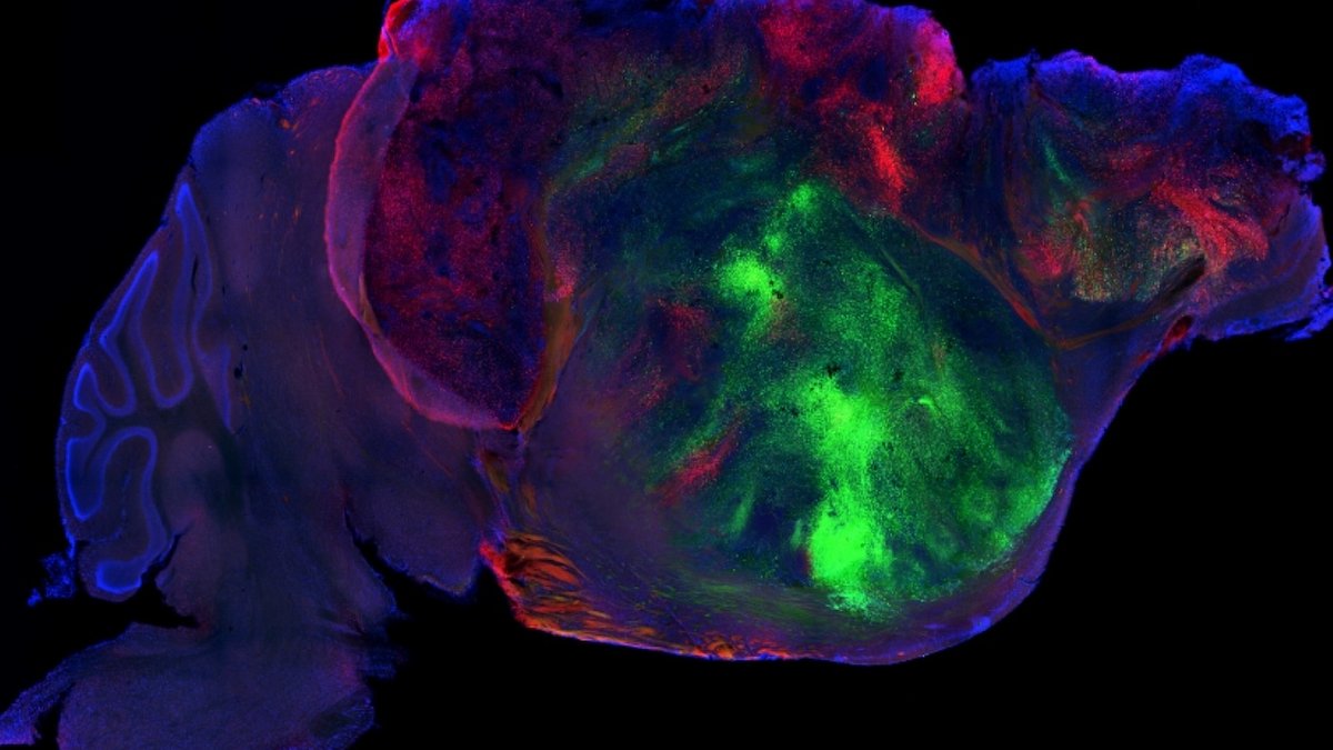
Pașca’s latest research, published in Nature, pushes these boundaries even further. Instead of transplanting into a normal mouse brain, his team created mice lacking most of their neocortex and hippocampus. These apallial mice are missing the dorsal and medial pallium—the regions that normally develop into those structures. The result is a brain with a large vacant space, primed to accommodate human cortical organoids.
Within three months, the human tissue filled the gap. It generated diverse cortical cell types, established long-range connections with the mouse nervous system, and developed organized electrical activity. The success rate was high: 86.2% of transplanted animals showed successful growth, with human-derived tissue comprising 91.9% of the cortical volume after three months. While the grafts did not form a perfect human cortex, lacking canonical lamination and full maturation, they contained a broad array of cell types, including astrocytes.
The additional space made a critical difference. Human neurons extended axons toward the superior colliculus, while mouse neurons from subcortical structures reached into the graft. Even the cervical spinal cord showed human-derived projections, a result not observed in prior transplants where the mouse cortex remained intact. The model also produced three times more layer 5 extratelencephalic (L5-ET) projection neurons, including cells resembling von Economo neurons (VENs), which are associated with frontotemporal dementia and other neuropsychiatric disorders.
Read Also: US approves new targeted cancer therapies
Pașca’s team demonstrated the model’s utility by studying hypoxic injury. When oxygen levels dropped, human grafts exhibited strong HIF1α activity, a stress response marker, while the adjacent mouse paleocortex showed no reaction. The microglia in the mouse brain responded to the injury only because the human cells were present. Pașca noted, “They’re reacting to the injury of human cells. But they’re not reacting to a hypoxic injury of the mouse cortex because there is essentially no mouse cortex there.”
The implications are significant: this system allows researchers to study human neural tissue within a living circuit, bridging the divide between cellular phenotypes and behavior. A hyperactive neuron in a dish does not guarantee seizures or EEG changes in a living system. But in xenocortical mice, those connections become observable. Pașca explained, “For many neuropsychiatric disorders, circuit and behavioral readouts are very important because psychiatric disorders are behaviorally defined.”
Developmental mismatches limit full integration
However, the model has constraints. Human neurons develop on their own timeline, reaching a mid-gestation equivalent after 24 weeks in culture, while the mouse nervous system matures much faster. Critical developmental windows in the mouse close before the human grafts reach maturity. Pașca noted that the cells are still progressing at their own pace, creating a hybrid system where human tissue grows but does not fully integrate into a mature cortical structure.
The apallial mice themselves revealed an unexpected insight. Despite lacking a cortex, these mice could still move, see, hear, and smell, though their gait and coordination were impaired. The nervous system had adapted, indicating that subcortical structures could compensate for missing cortical functions. Pașca stated, “It’s still not a fully formed human cortex, but it contains a large diversity of cortical cell types, including astrocytes.”
The work also raises ethical considerations as human neural grafts grow larger and more integrated. Pașca’s team consulted external committees and emphasized the need for proactive ethical oversight. For now, the focus remains on what this model can achieve, studying disease, testing treatments, and exploring the limits of neural development. The system is reproducible, though not straightforward. The mice can be generated using existing genetic tools, and organoid protocols are widely shared. Yet it is not a plug-and-play method, requiring live animals, human stem cells, and long-term experiments.
The model’s ability to generate von Economo neurons (VENs) in a rodent brain is particularly significant. These rare, large projection neurons are typically found only in humans and a few other large-brained mammals, such as elephants and whales. Their presence in the xenocortical grafts, verified through molecular markers and morphological features, opens new avenues for studying disorders linked to their dysfunction, including frontotemporal dementia and schizophrenia. Pașca’s team confirmed the cells’ identity using single-nucleus RNA sequencing, which matched their gene expression patterns to human VENs. The capacity to produce these neurons in a living system could help researchers test how their loss or dysfunction contributes to cognitive and behavioral symptoms.
Read Also: AI Enhances Aneurysm Detection but Increases False Alarms
Behavioral and Circuit-Level Readouts
Linking human neuron defects to mouse behavior
The most immediate practical application of the xenocortical model lies in its ability to connect human cellular phenotypes to observable behavior. Previous organoid systems allowed researchers to identify hyperexcitable neurons or abnormal calcium signaling, but these traits did not always translate into seizures, cognitive deficits, or other clinically relevant outcomes. The new model changes that. By transplanting patient-derived organoids, such as those from individuals with autism spectrum disorder or epilepsy, into apallial mice, scientists can now track whether cellular abnormalities lead to altered movement, sensory processing, or other behavioral changes. For instance, when human neurons in the graft became hyperactive, the mice displayed involuntary orofacial movements, suggesting a direct link from cellular dysfunction to motor output.
Electrophysiological recordings further demonstrated that human-derived activity could synchronize across large regions of the graft, producing coordinated bursts that aligned with the mice’s movements. This level of circuit organization was absent in earlier organoid models, where activity remained localized. The model also enables pathway-specific experiments: researchers can selectively activate or inhibit human neurons and observe the downstream effects on the mouse’s behavior. This could help distinguish between necessary and sufficient conditions for specific neural functions.
The model’s limitations persist due to the fundamental challenge: the mismatch between human and mouse developmental timelines. While human grafts develop at their own pace, reaching mid-gestation equivalence after 24 weeks in culture, the mouse nervous system matures far more rapidly. By the time the human tissue begins forming organized circuits, many of the mouse’s critical developmental windows have already closed. This prevents full synchronization, leaving gaps in functional integration. For example, the human grafts lack canonical cortical lamination, a hallmark of mature human cortex, because the cells lack structural guidance. Instead, they form clusters of related cell types, indicating a form of self-organization without external scaffolding.
Despite these constraints, the xenocortical model already provides an unprecedented opportunity to study human neural development in a living system. Pașca’s team has made the genetic tools and organoid protocols widely available, ensuring reproducibility. The mice, generated by crossing existing Emx1-Cre lines with immunocompromised strains, do not require novel genetic engineering. The human side of the system, growing cortical organoids from pluripotent stem cells, has also become more accessible, with Pașca’s lab training nearly 500 laboratories worldwide in organoid culture techniques.
However, the model is not a simple or rapid process. It demands long-term experiments, live animals, and precise handling of human stem cells, making it more complex than standard lab techniques like CRISPR. For now, its adoption will likely be limited to labs with expertise in both stem cell biology and animal models.
